-
 Human
Human
-
 Grade 9+


Electroencephalogram (EEG): See Your Own Brain Activity
Our brain is incredibly complex, yet measuring its electrical activity can be surprisingly accessible. In this experiment, we’ll record a type of brain signal called the electroencephalogram (EEG). You’ll learn how to spot the alpha wave—a rhythm first described by Hans Berger over 100 years ago.
About experiment

What Will You Learn?
- How to record EEG signals from your own scalp—no drilling required.
- Why EEG looks "wavey" and what it can (and can’t) tell us.
- The role of synchronous activity in generating detectable rhythms.
- How to measure the 10 Hz alpha wave and see it change with eyes open vs. closed.

Background
EEG measures the combined activity of millions of cortical neurons through electrodes placed on the scalp. Hans Berger, intrigued by a near-death incident that spurred thoughts of telepathy, discovered these rhythms in the 1920s. He noticed they change when eyes open or close. You’ll replicate his classic observation and decide what the patterns reveal about visual cortex activity.
Experiment
Pre-Frontal EEG Recording
Materials:
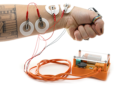
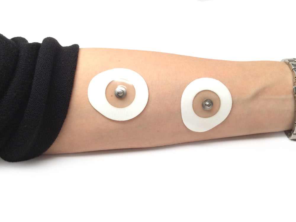
- Human SpikerBox
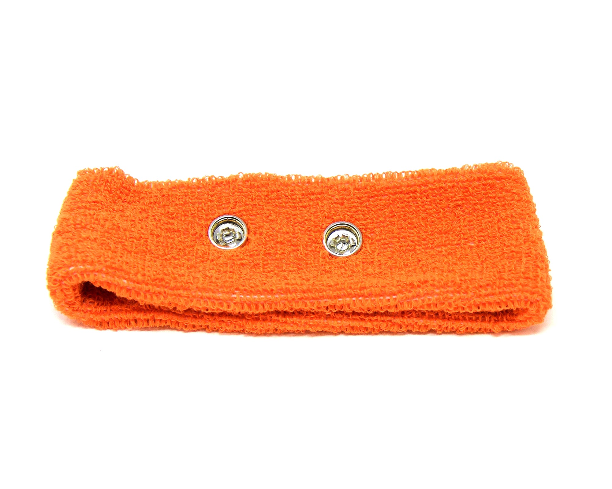
- Headband electrode + electrode gel
- EKG sticker for ground
Setup & Procedure:
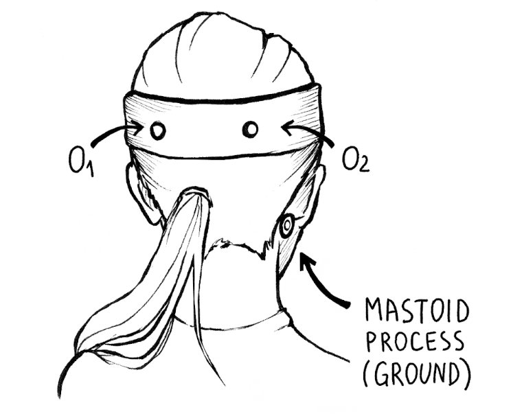
- Place headband so rivets touch skin above eyebrows.
- Ground electrode on mastoid behind ear.
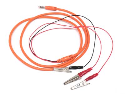
- Apply gel, clip leads (red to rivets, black to ground).
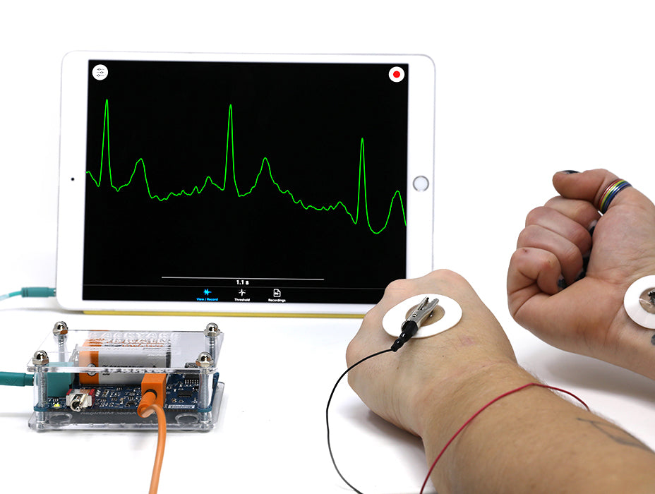
- Open SpikeRecorder. Adjust until you see low-amplitude waves.
- Think of colours, numbers, stressful vs. relaxing thoughts. Note if raw EEG shifts.
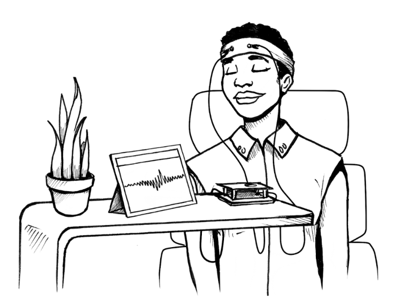
Occipital EEG Recording
Materials: Same kit as above.
Setup & Procedure:
- Rotate headband so rivets sit over occipital lobe.
- Gel through hair for solid contact.
- Record while alternating 10 s eyes open / 10 s eyes closed. Mark each switch.

Results & Analysis
Compare traces. With eyes closed, alpha waves (~10 Hz) usually strengthen and become more regular. Count peaks per second and measure amplitude. Discuss what this says about visual cortex engagement.
What do you need?
-
Related Products